Vicious Cycles
Before my exploration into visual arts and the advertising realm, I started my career in the scientific/medical field. A good chunk of my science career was dedicated to debunking the terrors of itch, inflammation, and pain — yes, that notorious reflex we experience during an unknowing attack by an obnoxious mosquito. But just on metaphorical steroids — when a combination of genetic factors, environmental stressors, and chemical allergens come into the attack for an uncontrollable itch, inflamed lesions, and often incessant pain. And it was our job to stop this vicious cycle of ruptured flesh inflicted by the over-enthusiastic scratching of that notorious itch! And did I mention how it can lead to leaky pustules that couldn’t be controlled with over-the-counter antihistamines (which primarily interact with a subset of receptors called H4R). Yes, itch like THIS was the kind of horror show that we played around in! With a top-notch international team of scientist hailing from the scientific research epicenters of Germany, Japan, and the USA, it appeared that Pruritus’ defunct cellular communication would finally be disarmed once and for all!
Pathway of pain and pruritic stimulation from the skin to central nervous system through peripheral nervous system. Infographic based on a scientific review article by my prior PI titled “Frontiers in Pruritus Research: Scratching the Brain for More Effective Itch Therapy” (nice title) published in the Journal of Clinical Investigation, 2006 (source).
Technicolor Dreams
There are design concepts used in formulating science research — one can argue that any form of experimentation is an art form. I especially enjoyed developing beautiful immunofluorescence or immunohistochemistry images of different tissues for markers we were looking to track, which provides insightful visual proof on your predictions. I've attached an image that we’ve produced for one of the projects I was working on involves pruritus, along with one created by a team at John Hopkins University that also found a neurological correlation between itch and pain. The purpose of this experiment was to see if our novel itch-inducing agonist, peptide Interleukin 31 (IL31), and it's corresponding receptor Interleukin 31 Receptor A (IL31RA), was somehow related to various known neuronal pathways for pain and inflammation signaling.
Immunofluorescence stained murine dorsal root ganglion (DRG) neurons of various pain-sensing neurons (red) and itch-sensing neurons (purple and yellow) triggered from the skin. Image credit: Xinzhong Dong et. al from John Hopkins University (source).
Getting Glowed-UP
In images, a, b, and c, the round structures are individual neurons in a section of mouse dorsal root ganglion (DRG), which is essentially a ball of neurons that acts as a relay station between the peripheral nervous system and the central nervous system. The dorsal horn of the spinal cord is also visualized (as seen in images d & e), which receives several types of sensory information from the DRGS including (aside from itch and pain) fine touch, proprioception, and vibration from the body. And more specifically, the various levels of laminae in the dorsal horn receives sensory projections from the body and coordinates the type of sensory information to be processed with the support of additional cell types (e.g interneurons). The sections have been labeled with fluorescent-labeled antibodies that bind to specific protein structures that they have been bioengineered to compliment — like glowing push-pins on a cork board except we're using a cross-section of tissue, and the pin only attaches to specific shapes of material.
(a,b,c) Immunofluorescence stained murine dorsal root ganglia (DRG) of itch-receptor IL31RA with various neuromarkers (TRPV1, IB4, and N52). Image credit: Ferda Cevikbas et. al. from University of California, San Francisco (source).
STOP it like it’s hot
At the far left column, you will see that our receptor of interest in red (IL31RA), which we believe directly modulates itch, hangs-out (or co-localised & appear yellow by image merge) on some of the same cells as transient receptor potential cation channel subfamily V member 1 (woo* that’s a typeful) labeled in green (or TRPV1/capsaicin receptor/vanilloid receptor for short). You're probably very familiar with the (arguably delicious) burning sensation of capsaicin when you bite into a jalapeño popper. This so happens to be correlated with endorphin and dopamine release via another molecule released called Substance P — talk about a foodie’s high! It’s been suspected that pain overrides some of the itch signals to reduce its rage - hence one’s natural reflex to scratch that itch.
Scientists have cleverly identified the receptor to these more specifically capsaicinoid molecules as the same ones that get turned on by hot temperatures of over 109˚F (43˚C). And have capitalized its use as the stereotypical an acute pain receptor to thermal-mechanical stimuli. On the same type of tissue, with two true positives are used to detect a specific subpopulation of neurons which include Lectin IB4 — a marker for non-peptidergic, unmyelinated sensory neurons, which is typically related to neuropathic pain, and modulates mechanical stimuli. The other is Neurofilament N52, which is a marker for neurons with myelinated axons that allow for fast responses, like pain transmission.
(d) Immunofluorescence stained murine dorsal horn of the spinal cord from control subjects (received intrathecal injection of vehicle) IL31RA and TRPV1, and (e) same procedure except from experimental subjects with TRPV1+ neurons ablated with an intrathecal injection of capsaicin. Image credit: Ferda Cevikbas et. al. from University of California, San Francisco (source).
A Nervous Breakdown
Based on images d and e, it appears that IL31RA is found on some of the same TRPV1+ neurons in the nervous system’s relay station (DRGs), and almost 100% coordination (co-localisation) in the dorsal horn of the spinal cord. This was further confirmed by ablating (basically burning away) all TRPV1+ neurons with a spinal flood (intrathecal injection) of capsaicin. However, this itch signaling pathway is only weakly related to a subpopulation of non-peptidergic, unmyelinated sensory neurons (N52), with almost no relation to myelinated neurons (IB4). So basically, IL-31 itch stimuli are strongly correlated with acute pain (TRPV1), but not part of our body’s fast-reacting response system (N52) or neuropathic pain pathways (IB4). When comes to modulation in the spinal cord, most of the IL31RA+/TRPV1+ neurons are found in laminae II which means they are involved in processing injury and inflammation and are concerned with pain sensation. And they probably do this with the help of excitatory interneurons that commonly occupy laminae II and III
Ferda Cevikbas, the principal researcher of IL-31 on skin disease, speaking at the 2011 International Conference on Itch in Tokyo, Japan (video source: Victoria Fong).
Recovery Pathways
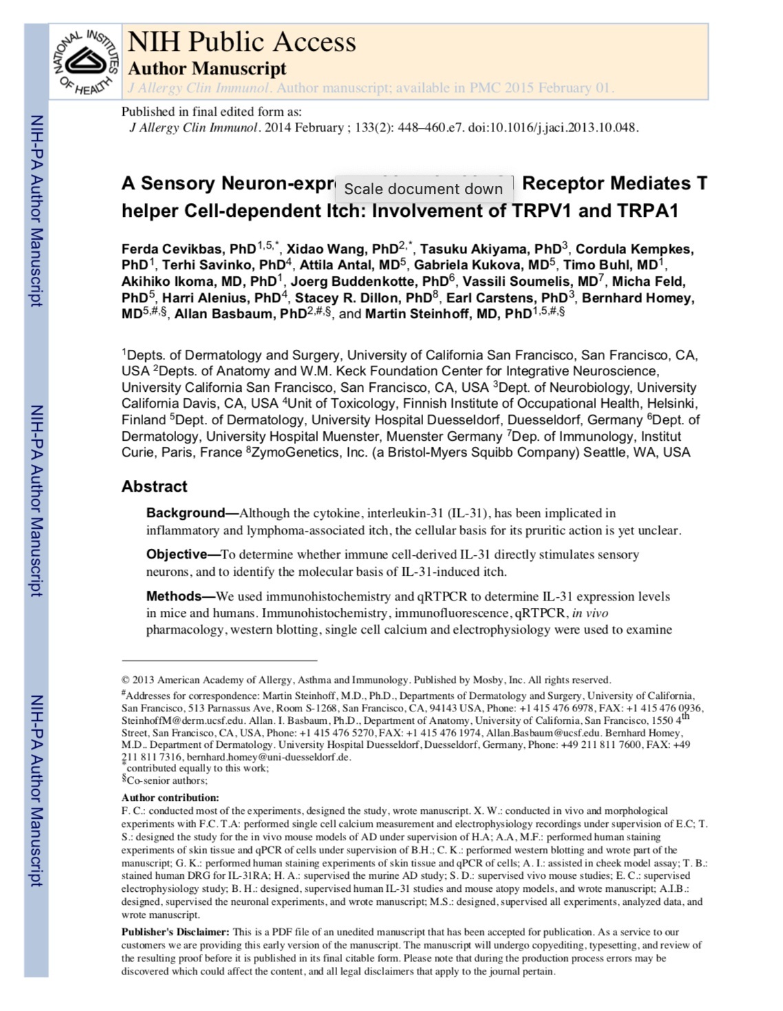
This is super interesting because this is visual proof of how acute pain and itch may have coordinated signaling functions through the TRPV1 pathway, and is experienced directly in your central nervous system by IL-31 cytokine signaling. Unfortunately, this means that IL-31/IL31RA related itch cannot be entirely treated by topical methods. But on a more optimistic note, there may be the potential of directly targeting its afferent neurons in the central nervous system, though it would only be possible if the treatment could somehow bypass the blood-brain barrier, e.g. in a compromised CNS caused by multiple sclerosis. Nonetheless, it’s still another scratch closer to helping us pave a pathway that can control antihistamine-resistant pruritus. Our publication “A Sensory Neuron- expressed Interleukin-31 Receptor Mediates T helper Cell-dependent Itch: Involvement of TRPV1 and TRPA1” further elaborates on the molecular basis of T-Helper immune cell-derived IL31 on itch, and the way this signaling differentiates from the classic pain pathways. I’ve also included an infographic that I’ve created on the general overview of these “Pruritic Pathways” that’s based on the publication.
Anyway, I hope I've tickled your … nervous system … with some itchy science (no hemorrhoids here, I promise)!
A general overview on the potential peripheral and central pathways for the novel itch molecule IL-31 based on my prior research lab’s publication: “Our publication “A Sensory Neuron-expressed Interleukin-31 Receptor Mediates T helper Cell-dependent Itch: Involvement of TRPV1 and TRPA1” (source).
Here is a framed photo of my lab group giving their best Matrix moment (or SnazzyFresh statement). This was given to me as a parting gift when I decided to leave the team to explore life beyond the lab bench. Thank you for everything that you have given to me— it was a stress at times, but mostly the best of times, and a blessing from the Universe to have been set up with you fine folks.
Update: Last edited in May 1, 2019


