Kidney Runner

OEA_WM anatomy_050919.png

EAST MEETS WEST


ON KIDNEY FUNCTION


How the Kidneys are the Root of Wellness
in Chinese and Western Medicine.

With pesticides and herbicides as a primary additive to our food systems; water pollution being a global catastrophe showing no slowing down; regular alcohol consumption consistently promoted as a social norm; the prescription of pharmaceuticals as the primary tool for resolving a chronic pain epidemic: I think it’s fair to say that the general public in the U.S. is pretty oblivious to how important our kidneys are for maintaining the homeostasis of our bodies. Not only does it function as the filtration system for the bodies’ perpetual production of potentially toxic waste products, it produces some of our bodies’ most important regulatory molecules like glucocorticoids for the infamous flight-fight-fright response (cortisol, adrenaline/epinephrine, and norepinephrine), your reproductive hormones (including androgens), and aldosterone for regulating blood pressure, to name a few. These molecules also happen to function as both neurotransmitters and hormones — that means these molecular messengers can essentially interact with any organ system of the body. In Traditional Chinese Medicine, the kidney is more of a description of the interrelated capabilities involved with the actual two physical organs. TCM practitioners also consider the kidney system the root of the energetic storage house of the body (or the “ming men”). In the infographics below, I’ve tied in some of the interrelating biochemical functions described by our Western Medical System with key descriptions from Traditional Chinese Medicine. The goal is to better understand why our kidneys should be considered one of the most important organ systems for indicating an imbalance in our bodies. Hopefully, it will inspire you to better take care of your magnificent pair. -VF

“If the water of the kidney cannot control the fire of the heart, the mind-spirit will become restless.”

The Renin-Angiotensin-Aldosterone System (RAAS) controls the blood pressure for fluid filtration and electrolyte balance through a network of checks-and-balances between the kidneys and the heart, though effects are experienced throughout the body. Hyperactivity of the RAAS system is also observed in depression. 

“THE Kidney storeS Jing or the Essence of Life — the substance that nourishes, fuels, and cools the body.”

The characteristics of strong Jing include prominent facial features, hearing, teeth, hair, and strong adrenals/kidneys. These traits share the same embryological origin as neural crest cells (NCCs) — a stem cell population with great abilities of self-organisation that undergo challenging cellular migrations to form the associated structures. Jing is both inherited (genetics) and acquired (food/water). 

“The Kidney is to dominate the regulation of Water Metabolism.”

The kidneys’ glomerular filtration removes excess fluid and waste products from the blood while playing a central role in the homeostasis of ions that are important for many biological functions (e.g. calcium, Vitamin D, etc.). 

“the kidney governs the bones, a substance born from water.”

The kidneys control the levels of calcium, Vitamin D/Calcitriol, phosphate, and cortisol, which are responsible for forming the crystalline structure of your bones. These compounds are also important for the regulation of central and peripheral mechanisms.

“The Kidney controls the reception and holding of the Qi to support the functions of the Lung.”

The regulation of acid-base equilibrium, modification of partial pressure of carbon dioxide and bicarbonate concentration, and the control of blood pressure and fluid homeostasis are all closely dependent on renal and pulmonary activities. 

 “The Kidney energy controls the sex drive and reproduction.”

Through the core energy center or “ming men”, the kidneys are depleted of “jing” during intercourse, and thus overexertion is warned against. We now understand that the kidneys are intimately connected to the adrenal glands, which release sex hormones (i.e. estrogen and testosterone) along with other neurohormones that regulate other functions like the stress response. The ovaries and testes also emerge from the primitive kidney during embryological development.

“The Spirit of the Kidney rules Willpower and the Survival Instinct.”

The kidneys produce many types of important hormones (histamine, serotonin, brain natriuretic peptide, adrenalin, norepinephrine, dopamine, etc.) that function universally as neurotransmitters, and are intimately connected with our state of being, our emotional and psychological health, and the seat of our primal state of fear.

I Gut A Feeling

Vagus Nerve.png

A GUT FEELING

FEAT. THE PAR2 IN YOU

The GI tract is the largest interface between you and the environment, and a primary influencer of your well-being, which is finally becoming increasingly understood as more research comes out (source). For the very first job that I landed fresh out of college, I was able to get my hands dirty (literally) in understanding the neurobiology of digestive disorders at a UCSF research lab (aka partaking in projects that required the dissection of mouse spinal cords and clearing of their intestines of poop). Oh the shit that I used to do… (cue punchline music*). Anyway, the founder of the lab had apparently done some major pioneering work on the role of this receptor, called Protease-activated Receptor 2 (PAR2 for short), in chronic pain associated with irritable bowel syndrome and inflammatory bowel disease. His team’s goal was to see if PAR2 could be exploited as a therapeutic target for chronic pain (source). Since then, I’ve developed a more holistic perspective on digestive health, but will always appreciate the impact of one problematic molecular mechanism on the whole. In homage to my time with digestive dysfunction, I’ve also created a karaoke video about the PAR2 in U in hopes of introducing an enjoyable way of learning about the science behind your well-being. -VF


An easy-to-digest overview of YOur Gut FEELINGS

SEROTONIN IS CONTROLLING

Besides being a critical processor of the nutritional requirements for this miraculous machinery we call our bodies, the human gastrointestinal system plays a major role in the “digestion” of your behavioral, emotional and physical capacities. This mechanism is monitored by a “second brain” which is officially known as the enteric nervous system (ENS): an expansive unit of over 500 million neurons (which is 0.5% the capacity of your brain and 5x that of the spinal cord), as well as a department of support cells. It relays information throughout your body from signaling pathways generated by but not limited to 30+ different cell types in the gut, molecules from digested nutrients (salts, fats, trace minerals, etc.), and the 100 trillion bacterial cells that support our digestion. Interconnected to the central nervous system (CNS) by the vagus nerve (and the lesser known prevertebral ganglia), the ENS lines the walls of the long tube of our gut, or alimentary canal, which measures about 30 feet end to end from the esophagus to the anus (source). Among one the critical communicators of the body, about 90% of the bodies’ serotonin reserves are manufactured by enterochromaffin (EC) cells in the gut with the support 2-6 pounds of bacterial flora, as discovered by a team at scientists at Caltech. Serotonin also regulates a whole entourage of biological processes including neurogenesis, cardiovascular function, intestinal motility, inflammation control, and bone mass accrual (source).

A BALANCING ACT

In Traditional Chinese Medicine, the organs, which aren’t delineated exactly the same as in Western anatomy, are responsible for regulating the whole spectrum of emotions that fluctuate throughout the day: the liver is the notorious wrangler of anger and frustration (most likely by filtering the blood of aggravating toxins); the energy center or “ming-men” of the kidneys monitors flare ups that cause fear in our hearts (which we now can partially prescribe to noradrenaline/adrenaline); our stomachs and pancreas process our worries in our immediate environments (by regulating blood sugar levels); and sadness is “digested’ by our lungs (ACE in regulating our blood pressure), skin (vitamin D for nervous system health) and large intestine (serotonin production). There’s also an additional non-physical digestive system called the triple burner residing over the head, chest, and thorax that controls all the organ relationships, and hence emotional balance — it is this heating system of the body/mind/spirit that regulates and distributes energy and water. Nowadays these functions can largely be attributed to the endocrine glands and hormonal distribution systems. It was also understood that the body maintains homeostasis (yin/yang) through processes that are analogous to the fundamental elements in our environment which, in general, are related to nutritional requirements (earth), power/organizational structure (metal), thermodynamics (fire), movement (water), and development (wood). How funny that practitioners have effectively applied this medical system for thousands of years without understanding the biochemical processes developed in Western Medicine, which have only gained traction over the last 200 years.

FOOD FOR FORM

One of the most overlooked aspects of the Western healthcare system is nutrition — in fact, most medical schools in the U.S. don’t even require their students to take a single nutrition course (source). In the ancient Indian medicine system of Ayurveda, health maintenance is entirely centered around nutrition, which include food, herbs and minerals (hence the culture’s delectable diversity of spiced delights). Prescriptions are made according to two main factors: your body makeup (prakruti), which consists of the distribution of three attributes (doshas), and six primary favours that support their functions. These three doshas, known as vata, pitta, and kapha, are correlated with the qualities of various biological energies that make up the body and mind. Interestingly enough, they have parallels with the mesomorph, ectomorph, and endomorph physiology defined in Western Medicine, which are influenced by the predominance of nervous tissue, bone mass, muscle, body fat percentage, rate of metabolism, etc. So it would make sense that specific foods would be more beneficial to someone with a tendency to gain weight (e.g. light salads that counteract the kapha dosha’s tendency for a sluggish metabolism) versus someone prone to weight loss and anxiety (e.g. sour foods for the vata dosha like oranges that contain vitamin C to promote healthy nervous system function). Your gut can even “taste” the macronutrient composition of the meal to trigger adequate responses for assimilating nutrients and eliminating waste products from your meals. Since Shirazi-Beechey’s pioneering work on the gut as a direct interface with the environment, researchers have found a specific distribution of receptors that detect sugars, fats, the amino acids (proteins), and bitter compounds in the GI tract — as well as all over the body including the lungs, CNS, testes, and skin (source). In fact, in Ayurveda clear skin is indicative of a healthy digestive system and overall balance of the body or three doshas (source).

ADDRESS THE STRESS

Normally a burst of cortisol is a healthy and necessary part of responding to a stressful situation through your ever-faithful fight-flight-freeze response. In fact this little glucocorticoid hormone has some major body-credentials that include: redistributing energy reserves like making your glutes more responsive to glucose (by thwarting the effect of insulin) when you’re running away from your problems; preventing you from passing out as that blood pressure ups the ante while you finally confront your demons; restrains any infected wounds from turning you into a swollen sack of flesh (for about 30 minutes) as a potent anti-inflammatory; and helps you selectively forget that the whole mess ever happened (memory consolidation). Your digestive system, on the other end, is curbed by the sympathetic nervous system for your “spidey senses” to operate at peak capacity — except for your liver which is on overdrive pumping out proteins to breakdown into glucose molecules for efficient fuel. All is well until stress becomes a chronic issue: it makes you grow a “beer belly” (from your brain’s cues to overeat and cortisol’s hand in fat cell production); you’re at a greater risk for Type II diabetes (from insulin resistance), a heart attack (constricted blood vessels), and autoimmune diseases (insulin resistance); your muscles start to waste away (dysregulation of the parasympathetic nervous system for nutrition processing); and you’re prone to a whole slew of mental health issues (e.g. insomnia, chronic fatigue syndrome, dementia, depression, etc.) (source).


The Role OF PAR2 in You

What is PAR2 and is it important in gastrointestinal health?

Protease-activated receptor 2 (PAR2), a receptor for inflammatory proteases, is a major mediator of neurogenic inflammation and pain. Injury and inflammation trigger the activation of proteases from the circulation, immune cells, and epithelial tissues that regulate cells by cleaving protease-activated receptors (PARs). Once activated, PARs regulate multiple pathophysiological processes, including inflammation, pain, homeostasis, and healing. PAR2 activation also promotes the central (brain, spinal cord) release of neuropeptides that activate second-order spinal neurons that transmit pain. These mechanisms contribute to painful inflammation of the intestine, pancreas, and joints (source).

How does PAR2 function on the molecular level?

PAR2 is a member of a family of four G protein-coupled receptors (GPCRs) - proteases released from injury and inflammation activate PAR2 on sensory nerve endings by cleaving it at specific sites within the extracellular N-terminal domains (outside of the cell). Proteases in circulation reveal tethered ligands that bind to and activate the cleaved receptors, such as the local release of these neuropeptides, which stimulate plasma extravasation (pus), infiltration of neutrophils, and vasodilation (neurogenic inflammation) (source).

What IS the role of PAR2 on the peripheral nervous system and skin?

Here is a brief video of the Principal Investigator (PI) that I used to work for at UCSF, Dr. Martin Steinhoff, briefly discussing how PAR2 also influences neuronal signaling and outgrowth in diseased skin. I shot this during the 2011 International Itch Conference in Japan, Tokyo which was organized by another scientific researcher I was working with at the time, Dr. Akihiko Ikoma.

A CLOSER LOOK at YOUR GUT LINING

The gut is far from a simple hollow tube — it’s full of cells that sense and respond to the contents, with ramifications for appetite, defense, and disease that researchers are just starting to uncover. This is an immunofluorescence stained cross-section of the lining of the small intestine that highlights the diversity of cells that constitute its structure: See the infographic “Protease-Activated Receptor (PAR2) Activation” that’s just below for a key to some the major cells found in the gut (and also pretty much throughout your body).

This is an immunofluorescence stained cross-section of the lining of the small intestine (image source).

THE CELLS IN YOUR GUT THAT carry PAR RECEPTORS

This is a summary of the pathophysiological effects of PAR agonists PAR1 and PAR2 are expressed by endothelial cells, epithelial cells, neurons, myocytes/smooth muscles, fibroblasts, and inflammatory cells. PARs trigger pathways related to inflammation, hyperalgesia, tissue repair and protection in these cell types.

Summary of the pathophysiological effects of PAR agonists PAR1 and PAR2.

I gut A feeling Feat. the par2 in u

As promised, here is the karaoke number on this multi-talented molecule that I've created in homage to my brief stint in GI research, but my lifelong respect for gut health. I used this research publication as a reference for the concepts and experiments conducted in PAR2 gut research, which was produced by a group of researchers that I used to work with. I’ve also included key below with definitions for the terms in the video that probably aren’t known by the typical lay-person. I hope you enjoy it!

P.S. This is not intended to downplay the importance of digestive diseases nor hurt anyone’s feelings — hopefully it will do the opposite, and maybe cause a slight cramp in your gut from a good guffaw.

KEY FOR "I Gut a Feelin’” Karaoke video

Basic Anatomy of the digestive system

Key for the basic identification of digestive organs for the "I Gut a Feelin’” karaoke video.

Cathepsin

Catheptic activity” (derived from the Greek word kathépsein, meaning to digest or to boil down) was first described in the gastric juice during the 1920s. Cathepsins are now classified based on their structure and catalytic type - the one used in mice experiments referenced in the research publication that I mentioned, Cathepsin S (for Serine), was found to excite the neurons in the colon.

C-FOS/C-Fos Immunohistochemistry Staining

C-Fos is a proto-oncogene that is expressed as a protein c-fos in some neurons upon depolarization (activation). It can be detected by immunohistochemical staining on tissue, such as activated nociceptive neurons in the spinal cord. We commonly used this tool to see if there was a central nervous system response (i.e. spinal cord) from mouse experiments on the gut.

Chronic Colitis

Colitis refers to the inflammation of the inner lining of the colon, which can be caused by several conditions including infection, Crohn’s disease, allergic reactions, and can have painful ulcers.

Colonic Air Balloon

A colonic balloon is just as it sounds, and used as part of an experimental technique for creating visceral gut pain in mice.

Confocal scope/Confocal Microscopy

Confocal microscopy is a type of microscopic imaging that uses lasers programmed to focus on a specific plane of tissue that’s been labeled with immunofluorescent markers. It’s used to create pretty, magnified images like the one included above.

Crohn’s/Crohn’s disease

Crohn’s disease is an inflammatory bowel disease that can cause severe abdominal pain, severe diarrhea, fatigue, weight loss, and malnutrition. I don’t have Crohn’s (as conveniently compatible with the lyrics), but I have known one person who’s successfully managed his symptoms with a combination of medications, vitamins, diet, exercise, and medicinal marijuana.

Gallstones

Gallstones are hardened deposits of the digestive bile, that can form within the gallbladder. They can cause pain, nausea, and vomiting if they block the ducts of the gallbladder on their way out. This is a more common condition in patients with Crohn’s disease.

MPO/Myeloperoxidase Assay

Myeloperoxidase (MPO) is a peroxidase enzyme found the most in neutrophil granulocytes (a type of white blood cell ) — basically in the presence of pus released during inflammation. Think about the times you used hydrogen peroxide to disinfect a scrape. The peroxidase in blood, cells, bacteria that break down the H2O2 into water and oxygen (which is responsible for the disinfectant properties against some bacterial strains).

The Itchy and Scratchy Show at UCSF

Scratching that Itch

Vicious Cycles

Before my exploration into visual arts and the advertising realm, I started my career in the scientific/medical field. A good chunk of my science career was dedicated to debunking the terrors of itch, inflammation, and pain — yes, that notorious reflex we experience during an unknowing attack by an obnoxious mosquito. But just on metaphorical steroids — when a combination of genetic factors, environmental stressors, and chemical allergens come into the attack for an uncontrollable itch, inflamed lesions, and often incessant pain. And it was our job to stop this vicious cycle of ruptured flesh inflicted by the over-enthusiastic scratching of that notorious itch! And did I mention how it can lead to leaky pustules that couldn’t be controlled with over-the-counter antihistamines (which primarily interact with a subset of receptors called H4R). Yes, itch like THIS was the kind of horror show that we played around in! With a top-notch international team of scientist hailing from the scientific research epicenters of Germany, Japan, and the USA, it appeared that Pruritus’ defunct cellular communication would finally be disarmed once and for all!

Pathway of pain and pruritic stimulation from the skin to central nervous system through peripheral nervous system. Infographic based on a scientific review article by my prior PI titled “Frontiers in Pruritus Research: Scratching the Brain for More Effective Itch Therapy” (nice title) published in the Journal of Clinical Investigation, 2006 (source).

Technicolor Dreams

There are design concepts used in formulating science research — one can argue that any form of experimentation is an art form. I especially enjoyed developing beautiful immunofluorescence or immunohistochemistry images of different tissues for markers we were looking to track, which provides insightful visual proof on your predictions. I've attached an image that we’ve produced for one of the projects I was working on involves pruritus, along with one created by a team at John Hopkins University that also found a neurological correlation between itch and pain. The purpose of this experiment was to see if our novel itch-inducing agonist, peptide Interleukin 31 (IL31), and it's corresponding receptor Interleukin 31 Receptor A (IL31RA), was somehow related to various known neuronal pathways for pain and inflammation signaling.

Immunofluorescence stained murine dorsal root ganglion (DRG) neurons of various pain-sensing neurons (red) and itch-sensing neurons (purple and yellow) triggered from the skin. Image credit: Xinzhong Dong et. al from John Hopkins University (source).

Getting Glowed-UP

In images, a, b, and c, the round structures are individual neurons in a section of mouse dorsal root ganglion (DRG), which is essentially a ball of neurons that acts as a relay station between the peripheral nervous system and the central nervous system. The dorsal horn of the spinal cord is also visualized (as seen in images d & e), which receives several types of sensory information from the DRGS including (aside from itch and pain) fine touch, proprioception, and vibration from the body. And more specifically, the various levels of laminae in the dorsal horn receives sensory projections from the body and coordinates the type of sensory information to be processed with the support of additional cell types (e.g interneurons). The sections have been labeled with fluorescent-labeled antibodies that bind to specific protein structures that they have been bioengineered to compliment — like glowing push-pins on a cork board except we're using a cross-section of tissue, and the pin only attaches to specific shapes of material.

(a,b,c) Immunofluorescence stained murine dorsal root ganglia (DRG) of itch-receptor IL31RA with various neuromarkers (TRPV1, IB4, and N52). Image credit: Ferda Cevikbas et. al. from University of California, San Francisco (source).

STOP it like it’s hot

At the far left column, you will see that our receptor of interest in red (IL31RA), which we believe directly modulates itch, hangs-out (or co-localised & appear yellow by image merge) on some of the same cells as transient receptor potential cation channel subfamily V member 1 (woo* that’s a typeful) labeled in green (or TRPV1/capsaicin receptor/vanilloid receptor for short). You're probably very familiar with the (arguably delicious) burning sensation of capsaicin when you bite into a jalapeño popper. This so happens to be correlated with endorphin and dopamine release via another molecule released called Substance P — talk about a foodie’s high! It’s been suspected that pain overrides some of the itch signals to reduce its rage - hence one’s natural reflex to scratch that itch.

Scientists have cleverly identified the receptor to these more specifically capsaicinoid molecules as the same ones that get turned on by hot temperatures of over 109˚F (43˚C). And have capitalized its use as the stereotypical an acute pain receptor to thermal-mechanical stimuli. On the same type of tissue, with two true positives are used to detect a specific subpopulation of neurons which include Lectin IB4 — a marker for non-peptidergic, unmyelinated sensory neurons, which is typically related to neuropathic pain, and modulates mechanical stimuli. The other is Neurofilament N52, which is a marker for neurons with myelinated axons that allow for fast responses, like pain transmission.

(d) Immunofluorescence stained murine dorsal horn of the spinal cord from control subjects (received intrathecal injection of vehicle) IL31RA and TRPV1, and (e) same procedure except from experimental subjects with TRPV1+ neurons ablated with an intrathecal injection of capsaicin. Image credit: Ferda Cevikbas et. al. from University of California, San Francisco (source).

A Nervous Breakdown

Based on images d and e, it appears that IL31RA is found on some of the same TRPV1+ neurons in the nervous system’s relay station (DRGs), and almost 100% coordination (co-localisation) in the dorsal horn of the spinal cord. This was further confirmed by ablating (basically burning away) all TRPV1+ neurons with a spinal flood (intrathecal injection) of capsaicin. However, this itch signaling pathway is only weakly related to a subpopulation of non-peptidergic, unmyelinated sensory neurons (N52), with almost no relation to myelinated neurons (IB4). So basically, IL-31 itch stimuli are strongly correlated with acute pain (TRPV1), but not part of our body’s fast-reacting response system (N52) or neuropathic pain pathways (IB4). When comes to modulation in the spinal cord, most of the IL31RA+/TRPV1+ neurons are found in laminae II which means they are involved in processing injury and inflammation and are concerned with pain sensation. And they probably do this with the help of excitatory interneurons that commonly occupy laminae II and III

Ferda Cevikbas, the principal researcher of IL-31 on skin disease, speaking at the 2011 International Conference on Itch in Tokyo, Japan (video source: Victoria Fong).

Recovery Pathways

This is super interesting because this is visual proof of how acute pain and itch may have coordinated signaling functions through the TRPV1 pathway, and is experienced directly in your central nervous system by IL-31 cytokine signaling. Unfortunately, this means that IL-31/IL31RA related itch cannot be entirely treated by topical methods. But on a more optimistic note, there may be the potential of directly targeting its afferent neurons in the central nervous system, though it would only be possible if the treatment could somehow bypass the blood-brain barrier, e.g. in a compromised CNS caused by multiple sclerosis. Nonetheless, it’s still another scratch closer to helping us pave a pathway that can control antihistamine-resistant pruritus. Our publication “A Sensory Neuron- expressed Interleukin-31 Receptor Mediates T helper Cell-dependent Itch: Involvement of TRPV1 and TRPA1” further elaborates on the molecular basis of T-Helper immune cell-derived IL31 on itch, and the way this signaling differentiates from the classic pain pathways. I’ve also included an infographic that I’ve created on the general overview of these “Pruritic Pathways” that’s based on the publication.

Anyway, I hope I've tickled your … nervous system … with some itchy science (no hemorrhoids here, I promise)!

A general overview on the potential peripheral and central pathways for the novel itch molecule IL-31 based on my prior research lab’s publication: “Our publication “A Sensory Neuron-expressed Interleukin-31 Receptor Mediates T helper Cell-dependent Itch: Involvement of TRPV1 and TRPA1” (source).

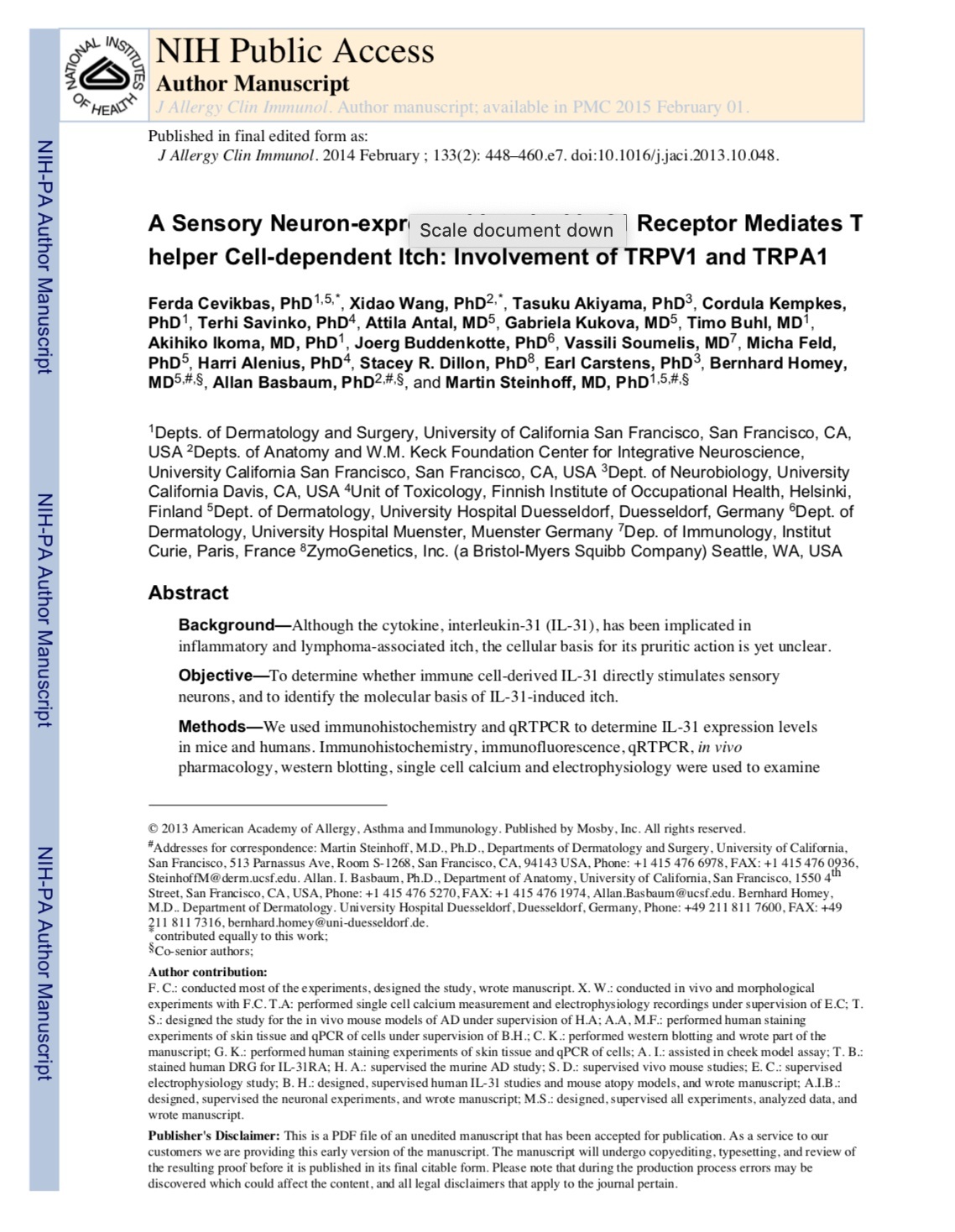
The research publication that most of this article is based on that I worked on with a few of the radest scientists ever (source).

The research publication that most of this article is based on that I worked on with a few of the radest scientists ever (source).

An in-depth review on the neurobiology of itch written by one of the post-doctorates and PI from my lab (source).

An in-depth review on the neurobiology of itch written by one of the post-doctorates and PI from my lab (source).

Here is a framed photo of my lab group giving their best Matrix moment (or SnazzyFresh statement). This was given to me as a parting gift when I decided to leave the team to explore life beyond the lab bench. Thank you for everything that you have given to me— it was a stress at times, but mostly the best of times, and a blessing from the Universe to have been set up with you fine folks.

Update: Last edited in May 1, 2019